

Skin cancer is one of the most common cancers worldwide, and melanoma remains one of its most dangerous forms. But when detected early, the survival rate is extremely high. This is why mole mapping technology has become one of the most important advances in modern dermatology. It transforms skin cancer care from reactive treatment to proactive prevention and provides both clinicians and patients with a powerful tool for long-term monitoring of skin health.
What is Mole Mapping?
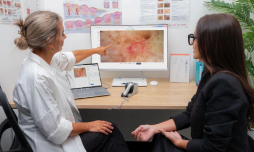
Mole mapping is a sophisticated digital imaging process that creates a detailed record of all moles and skin lesions on a patient’s body. It combines total body photography with close-up dermoscopic imaging to produce a complete visual map of the skin that can be reviewed, stored and compared over time.
How does Mole mapping work?
Unlike standard skin testing, which relies on visual inspection at a single point in time, the mole map creates a permanent digital baseline. Every injury is documented, measured and recorded so that any future changes, no matter how subtle, can be quickly identified and acted upon. This is not just a picture of sick skin today. This is a growing clinical record that becomes more valuable with each visit.
In the first phase, a full-body imaging session takes standardized, high-resolution images of the patient’s entire body surface from multiple angles. Many clinics perform this step using a system such as the MoleMax HD trolley, which is designed for easy mobility and comes with an integrated Windows PC, allowing the clinician to move all imaging settings between consulting rooms without disrupting workflow. Others rely on a common body image setup, such as PhotoMax Pro Suitewhich pairs a high-resolution SLR camera with an optional body-mapping stand for continuous, full-body recording session after session. This gives the clinician a complete overview of all visible moles and lesions throughout the body.
In the second stage, a dermatoscope is used to take close-up dermoscopic images of individual lesions that appear suspicious or require closer monitoring. These detailed images reveal the internal structures of each lesion at a higher resolution than what is visible to the naked eye during a routine skin examination.
In the third stage, all images are stored in a digital patient record and connected to a special software platform. At each subsequent visit, the new images are compared side-by-side with the previous images. This allows doctors to detect any changes in size, shape, color or texture with accuracy and confidence, making it easier to spot early warning signs before they turn into something serious.
Benefits of mole mapping for patients and clinicians:
Mole mapping brings significant benefits to both sides of the boardroom.
For clinicians, it significantly improves diagnostic accuracy by providing reliable visual reference over multiple visits. It reduces the risk of missing subtle changes in lesions that may not be apparent during standard visual examination. It also supports reliable clinical decision-making and helps clinicians avoid unnecessary surgical resections by monitoring borderline lesions over time rather than removing them prematurely. This leads to better patient outcomes and a more efficient clinical workflow.

For patients, mole mapping provides real reassurance and peace of mind. Knowing that every lesion on their body is closely monitored gives patients confidence in the long-term management of their skin health. It also encourages regular follow-up visits, which is important for early detection. High-risk patients in particular benefit greatly from having a complete and continuously updated digital record of their skin, giving them an active role in their own care.
Mole Mapping vs Traditional Skin Testing
A traditional skin exam by a doctor involves a visual examination of the skin, sometimes with the help of a handheld dermatoscope. While this approach has clear value, it also has limitations that become more important over time.
A traditional skin test shows what the lesion is at a given time. It relies on clinical notes and memory to track changes between visits, making it difficult to detect slow and gradual development in lesions. There is no permanent visual record that can be objectively compared at future appointments, which means that subtle but significant changes can be missed.
Mole mapping directly addresses all of these limitations. It replaces subjective recall with objective numerical comparison. It ensures that nothing is overlooked on the entire surface of the body. And it creates a growing archive of a patient’s skin history that reinforces clinical value with every appointment. In simple terms, a traditional skin test tells you what the current lesion is. A commodity map tells you how it has changed over time and whether that change requires action.
Who should review the product map?
Mole mapping is especially important for patients at high risk of melanoma. These include patients with multiple moles, a personal or family history of melanoma, fair skin that burns easily, a significant history of sunburn or sunburn, or a previous diagnosis of an atypical or dysplastic nevi.
However, mole mapping is not just for high-risk patients. Many people who do not have specific risk factors choose mole mapping as part of a proactive approach to their long-term skin health. As awareness of skin cancer continues to increase worldwide, more patients are seeking this level of monitoring as a standard part of their health care routine rather than waiting for problems to arise.
Frequently asked questions
How long does a product mapping session take?
A complete mole mapping session takes 30 to 60 minutes, depending on the number of documented lesions and the complexity of the patient’s skin profile.
How often should asset mapping be done?
For most patients, a mole map once a year is recommended. High-risk patients are advised to attend more frequently, such as every six months, based on their clinical profile and findings from previous sessions.
Is mole mapping painful or invasive?
No. Mole mapping is completely non-invasive and painless. It only involves photography and dermoscopic imaging, without any discomfort for the patient at any stage of the process.
Can a mole map detect all types of skin cancer?
Mole mapping is primarily intended for the detection and monitoring of melanoma and pigmented lesions. It is most effective when used as part of a broader skin cancer screening program that includes a complete clinical examination.
What technology is used in commodity mapping?
State-of-the-art mole mapping systems use high-resolution digital cameras, advanced dermoscopy equipment, and specialized patient management software. MoleMax Systems offers a comprehensive suite of mole mapping solutions, including the MoleMax HD PRO and MoleMax Plus software platforms, trusted by dermatologists and skin cancer clinics in more than 70 countries.
Is the mole map covered by health insurance?
Coverage varies by country and specific health insurance policy. It is recommended that you check directly with your provider or clinic about coverage and out-of-pocket costs.
Conclusion
Mole mapping technology is more than just an improvement on traditional skin testing. It represents a fundamental shift in how skin cancer care is delivered, moving from single-point assessment to continuous, data-driven monitoring. For dermatology and skin cancer practices, investing in mole mapping technology means offering a higher standard of care, increasing diagnostic confidence, and building a practice reputation based on clinical excellence.
MoleMax Systems offers a complete suite of mole mapping solutions designed for dermatology and skin cancer practices of all sizes. With more than 20 years of experience and a global network of more than 3,000 physicians in 70 countries, MoleMax is a trusted practice partner, ready to lead the way in modern skin cancer care.
Book for free 15 minute online show today and learn how MoleMax mapping technology can transform your practice.
- About the Author
- Latest news

Our editorial team are writers and experts in their field. Their views and opinions are not necessarily those of Wellbeing Magazine. If you are under medical supervision, talk to your doctor or therapist before following the advice and recommendations in this article.