

Over a year ago, I wrote a post about how trauma is “not just in your head.” Here, I follow it up with supporting evidence. I have devoted much of my life to trauma healing using emotion-focused therapy EMDR treatments. And I was constantly wondering how and why it works.
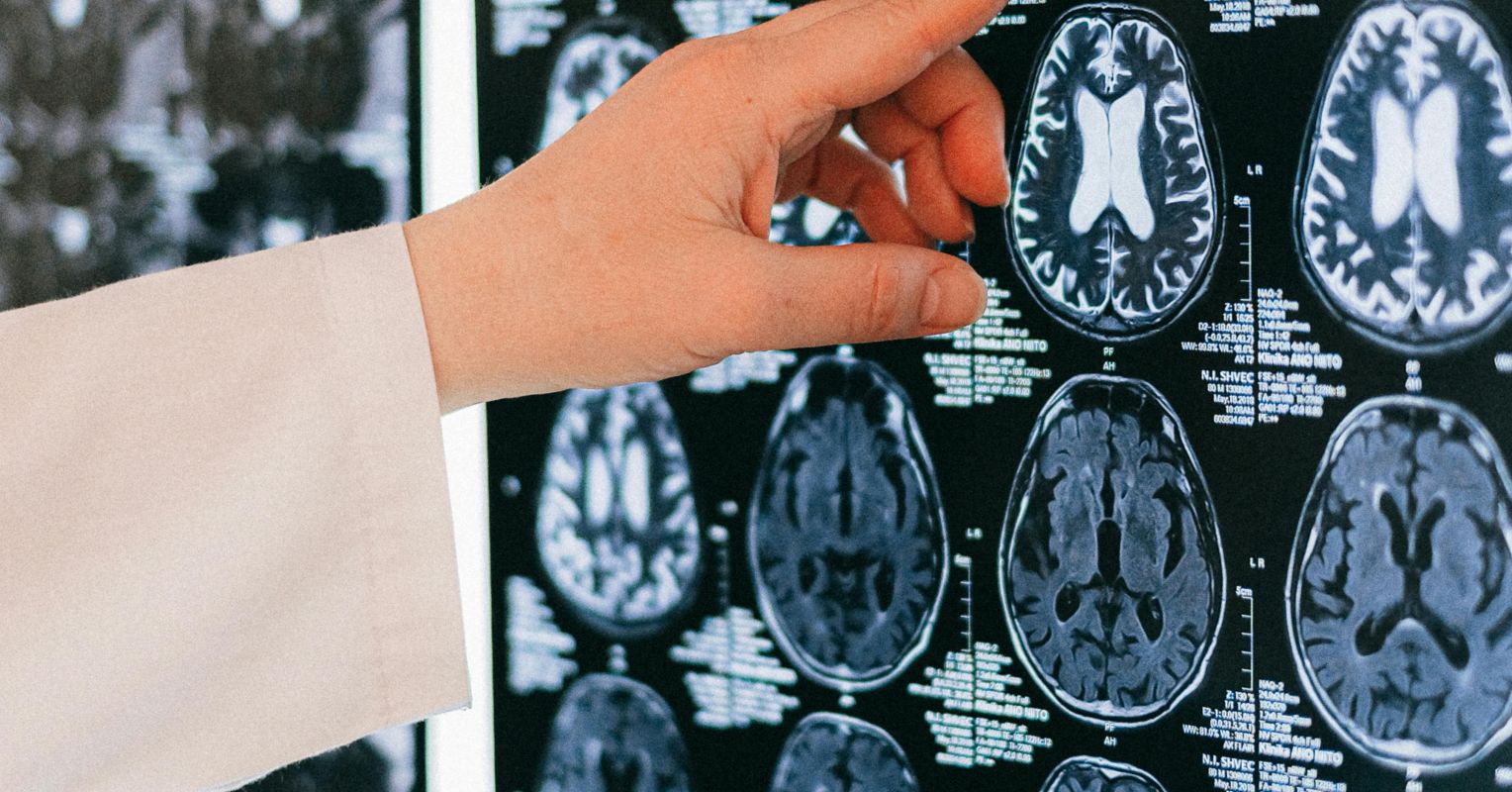
I also see versions of brain diagrams Social media constantly; A colorful infographic showing how trauma affects key areas of the brain. And while I tend to oversimplify mental health notesThis is really a very important thing. The neurology of trauma is well-documented, enlightening, and validating for survivors and their loved ones. I’m sure it’s deeply comforting and humanizing for any survivor who’s ever wondered: What happened to me? Or, why can’t I experience this?!
Here I summarize the four most commonly damaged brain regions and their connection to people seeking healing from real pain (as I mentioned in a previous post, “trauma” comes from the Latin word for “wound”).
1. The overstimulated amygdala: your brain’s faulty alarm system
For me, the most useful way to think about the amygdala is as your brain’s primary threat detection system. In the best case scenario, it fires when there’s real danger, helps you respond effectively, and returns to action once the threat is gone. During trauma (defined by one’s reaction to the event, not the event itself), this system not only stays “on,” but I would argue that the brain’s gear that keeps it “on” metaphorically clicks and locks it into “on,” despite the best intentions of cranking the dial so it can move fluidly depending on current threats. Functional studies consistently show that PTSD Patients show increased activation of the amygdala in response to threatening signals, with reduced activity in prefrontal regions that normally suppress it (Kredlow et al., 2022; Etkin & Wager, 2007). The result of a nervous system always looking for danger (and the like confirmation biasit seeks information that confirms an existing belief). It takes risk into account and therefore finds it even when there is none or the risk is minimal.
Therefore, trauma survivors are not dramatic when they are startled by a lightly pressed door, freeze when touched unexpectedly, or when they cannot rest safely in an objective environment. That being said, their brains are neurobiologically hard-wired to stay alert (even when they don’t need to be alert). As I’ve written about before, this brain bug isn’t a character flaw; it’s a sensitive alarm system that has saved their life one or more times and didn’t get the memo (and/or doesn’t trust the memo) that the threat has passed.
2. Damaged prefrontal cortex: When rationality goes offline
For me, the most useful way to think about the prefrontal cortex (PFC) is as your brain’s conductor or CEO. Its function is rational thinking, decision makingstrategy, tactics and most critically, evaluation and regulation of emotional responses from the amygdala. In victims, this control is violated. Research in Neuropsychopharmacology describes a well-supported model in which the ventromedial (“ventro” refers to the front and “medial” refers to the middle in brain anatomy) PFC, which normally controls and inhibits the amygdala, becomes hypoactive when the amygdala above becomes hyperactivity. This imbalance makes it much more difficult to regulate fear. (Kredlov et al., 2022).
This is why trauma healing is not just about thinking differently, a misconception I hear often even from health and mental health professionals. I repeat: You cannot take your logic out of your nervous system in an overloaded state. When the PFC goes offline under stressability to think rationally, perspectiveand emotional management all take a significant hit. It also explains why traditional discoursetherapy alone, although valuable, may be ultimately insufficient for many sufferers because it may not directly address the body’s and brain’s alarm systems.
3. Inactivated Broca’s area: Why it is impossible to “put it into words”.
Have you ever had the experience of knowing what you were feeling but not being able to find the right or appropriate words? I know I have. It’s not laziness or avoidance, it’s neurobiology. Our lived experiences and the resilience and richness of our emotional lives can transcend the boundaries of language, especially for trauma survivors.
Broca’s area, located in the left inferior (inferior) frontal gyrus (one of the brain’s main communication centers), is central to language production and labeling experience (especially emotional language). Neuroimaging research has consistently shown that Broca’s area is activated during the onset of trauma symptoms in PTSD (Hull, 2002), particularly at a key time when the amygdala is hyperactive (Rauch et al., 2006; Hull, 2002). One review described this deactivation as a possible explanation for trauma survivors’ difficulties in putting words to their feelings and experiences, which are essentially silenced when their traumatic memories are activated (Hull, 2002). This is why in EMDR therapy, we often ask the person what they are verbally observing because they are experiencing rapid eye movement trauma.
For me, this has profound clinical implications. It helps to understand why asking someone in their trauma state to talk in a highly active state can feel and be almost neurologically impossible. This is also a compelling argument for body-based and sensitive techniques such as EMDR (and somatic therapy) that do not require the client to verbally communicate that they are focusing on healing trauma (Shapiro, 2017). The brain can’t always put it into words because of the stress of the trauma, and so it often shuts down, like a computer running too many programs at once (or a computer overheating from hot air), until it’s forced to restart or shut down.
4. Shrinking hippocampus: When past and present collapse
The hippocampus is important for contextualization and formation memoryhelps us record experiences in a coherent timeline with clear markers of then and now. If you have a MacBook, you can think of it as a “Search” of folders that are basically displayed in chronological order. In PTSD, hippocampal volume reduction is one of the most consistently repeated neurobiological findings in the literature that I have seen. In fact, hippocampal volume can differentiate PTSD patients from trauma controls who have not developed the disease (Zilcha-mano et al., 2023).
This means vulnerable memories often fade away anchor in time. They do not feel that the memory of something that happened in the past but something still happens. A smell, a sound, a particular quality of light can transport a trauma victim into a full-body experience that feels present. This is not a hallucination; it is the hippocampus that cannot stamp the memory with “past” and “safe”. Fortunately, research also shows that the volume of the hippocampus can increase with successful treatment, and there is real hope / evidence that these changes are not permanent (Zilcha-mano et al., 2023).
Putting it together
These findings are why I gently but persistently push back when someone suggests that trauma is “all in your head” or that survivors should go it alone in recovery. These four brain regions: amygdala, prefrontal cortex, Broca’s area, and hippocampus are measurably altered in affected patients. Suffering is biological, somatic, physiological and emotional, not just mental. And the most effective techniques for trauma, such as EMDR, somatic experience, and emotionally focused therapy, among others, work precisely because they embrace these neurobiological realities, rather than trying to talk around them.
Understanding brain changes after trauma is not only intellectual but also validating. That experience from “What’s wrong with me?” repeats. to “It happened to my brain.” This change alone is important to me in healing progress.
To find a therapist, please visit List of current psychological therapies.